- info@kingsdiagnosticimaging.com
- Saint Charles Road Before Attaesibi Hotel
- Mon - Fri: 8:00 am - 6:00 pm
Gallery Posts


Our personal motivation consists of enhancing and improving patients’ experience in a sustainable manner. This is captured in our motto/slogan:
‘Legacy for Excellence’.
| Mon - Sat: | 8:00 am - 6:00 pm |
| Sunday | 11:00 am - 4:00 pm |
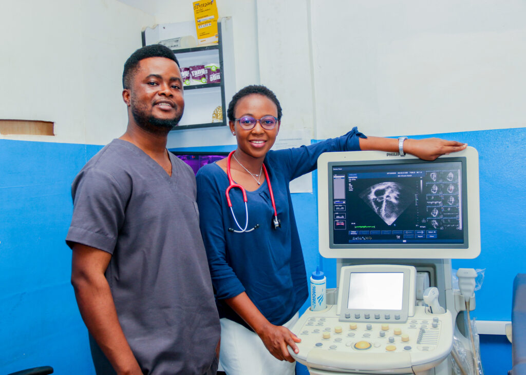
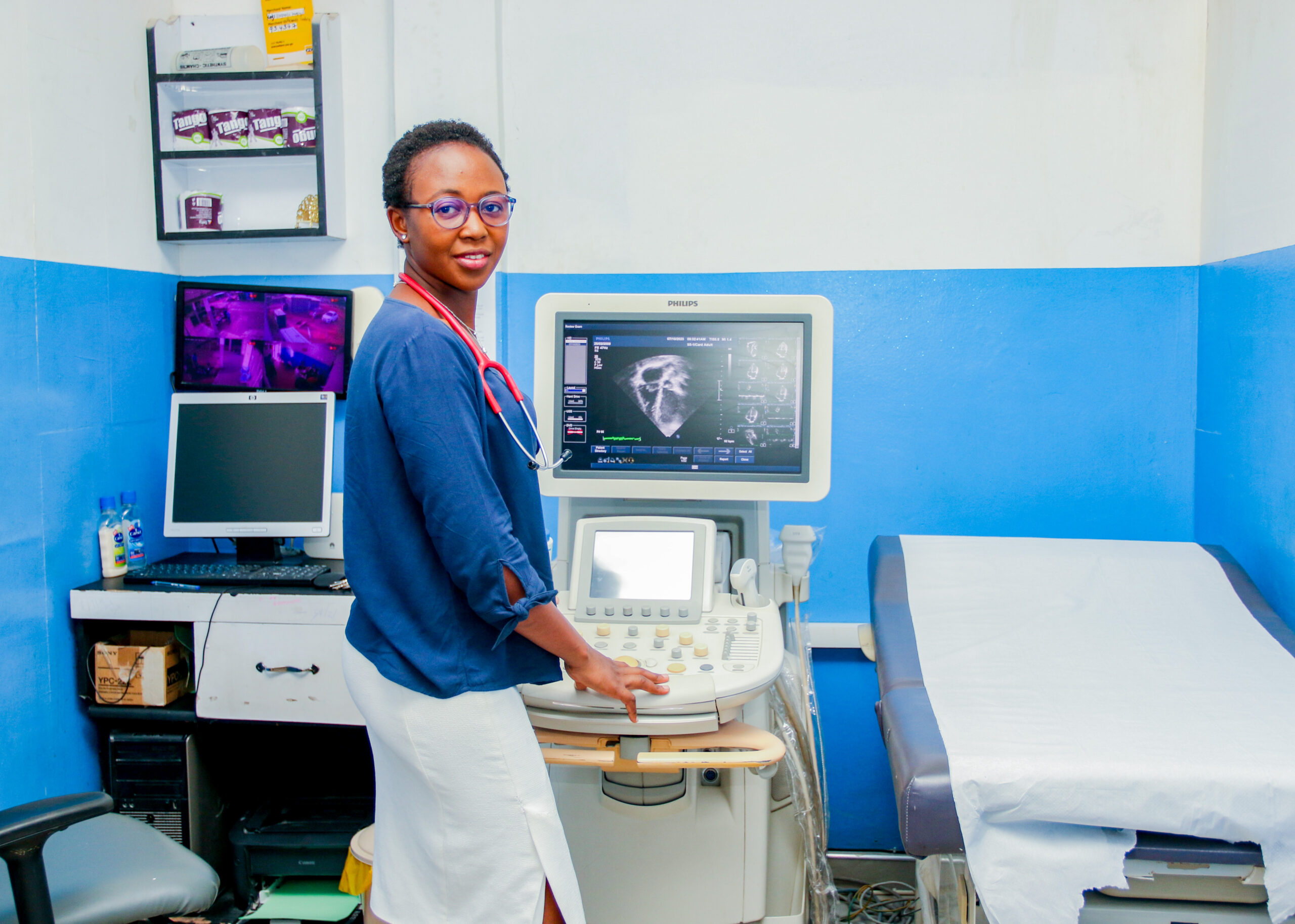
Women’s healthcare in the context of obstetric imaging and gynecological imaging involves the use of advanced medical imaging techniques to diagnose, monitor, and treat conditions specific to the female reproductive system.
Obstetric imaging refers to the use of various medical imaging techniques to monitor the health and development of a fetus during pregnancy and to assess the health of the mother’s reproductive organs. These imaging techniques play a crucial role in prenatal care and can help healthcare providers detect and diagnose potential complications or abnormalities.
Here are some common obstetric imaging techniques:
Gynecological imaging involves the use of various medical imaging techniques to visualize and assess the female reproductive system, including the uterus, ovaries, fallopian tubes, cervix, and surrounding structures. These imaging modalities are essential for diagnosing and monitoring gynecological conditions and guiding appropriate treatment. Here are some key aspects of gynecological imaging:
Transvaginal Ultrasound: Transvaginal ultrasound is a common imaging technique in gynecology. It involves inserting a small ultrasound probe into the vagina to obtain detailed images of the pelvic organs. Transvaginal ultrasound is useful for assessing the uterus, ovaries, and detecting conditions such as ovarian cysts, fibroids, and abnormalities in the endometrial lining.
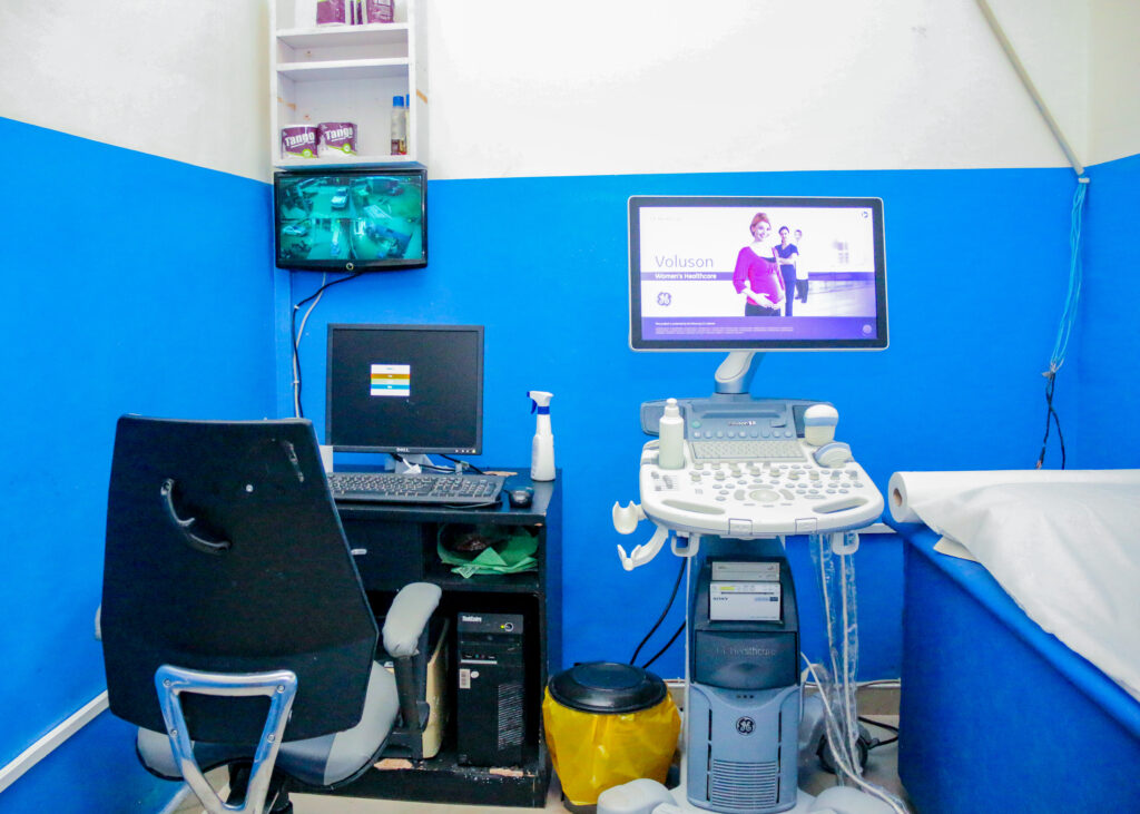
Pelvic Ultrasound: Pelvic ultrasound is performed externally on the abdomen and provides an overview of the pelvic area. It is used to evaluate the size, shape, and position of the uterus and ovaries, as well as to detect pelvic masses or structural abnormalities.
Hysterosonography: Hysterosonography, also known as saline infusion sonography, involves injecting saline into the uterine cavity while performing a transvaginal ultrasound. This technique is used to visualize the uterine lining and identify abnormalities such as polyps, fibroids, or adhesions.
Hysterosalpingography (HSG): HSG is a specialized X-ray procedure in which contrast dye is injected into the uterus and fallopian tubes to assess their patency. It is often used to investigate infertility and to check for blockages in the fallopian tubes.
3D and 4D Ultrasound: Advanced ultrasound techniques, such as 3D and 4D ultrasound, can provide three-dimensional images and real-time video of the pelvic structures, enhancing the diagnostic capabilities and visualization of gynecological conditions.
Our personal motivation consists of enhancing and improving patients’ experience in a sustainable manner. This is captured in our motto/slogan: ‘Legacy for Excellence’.


Copyright 2023 All Rights Reserved