- info@kingsdiagnosticimaging.com
- Saint Charles Road Before Attaesibi Hotel
- Mon - Fri: 8:00 am - 6:00 pm
Gallery Posts


Our personal motivation consists of enhancing and improving patients’ experience in a sustainable manner. This is captured in our motto/slogan:
‘Legacy for Excellence’.
| Mon - Sat: | 8:00 am - 6:00 pm |
| Sunday | 11:00 am - 4:00 pm |
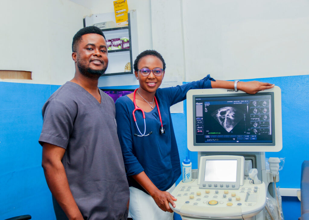
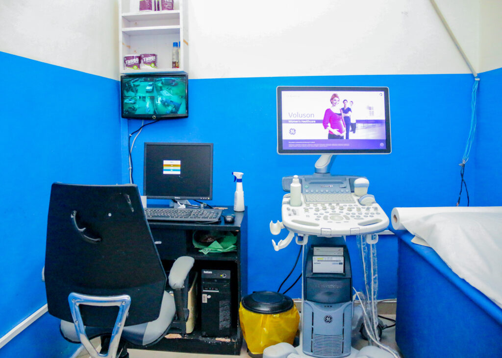
An abdominopelvic ultrasound, often referred to as an abdominal-pelvic ultrasound or simply an abdominal ultrasound, is a medical imaging procedure that uses high-frequency sound waves to create detailed images of the organs and structures in the abdomen and pelvis. This non-invasive and painless imaging technique is commonly used for diagnostic purposes to evaluate various conditions and diseases in these regions of the body. Here’s what you need to know about abdominopelvic ultrasound:
Purpose:
Abdominal Evaluation: An abdominopelvic ultrasound is used to assess the abdominal organs, including the liver, gallbladder, pancreas, spleen, kidneys, and aorta. It can detect abnormalities such as gallstones, liver cysts, or tumors.
Pelvic Examination: In addition to the abdominal organs, this type of ultrasound also provides a view of the female reproductive organs (uterus and ovaries) and the male bladder and prostate. It is used to diagnose conditions like ovarian cysts, uterine fibroids, and prostate enlargement.
Pregnancy Monitoring: Abdominopelvic ultrasounds are employed during pregnancy to monitor the development of the fetus, assess the placenta, and check for any potential complications.
Procedure:
Preparation: Depending on the specific reason for the ultrasound, you may be asked to fast for several hours before the procedure, especially if the examination focuses on the abdominal organs. Fasting helps improve the visibility of certain structures, like the gallbladder.
Positioning: You will typically lie on your back on an examination table. A clear, water-based gel is applied to the area being examined, which helps transmit sound waves and allows the ultrasound transducer (a handheld device) to move smoothly over your skin.
Scanning: The ultrasound technician (sonographer) moves the transducer over the abdominal and pelvic areas while observing a real-time image on the ultrasound monitor. They may ask you to change positions or hold your breath briefly to obtain better views of specific organs or structures.
Recording Images: The sonographer captures still images or records video clips of the areas of interest. These images are saved for review by a radiologist or healthcare provider.
Post-Procedure:
After the exam, you can usually resume your normal activities immediately. There are no radiation or significant side effects associated with an abdominal-pelvic ultrasound.
Results:
The recorded images and findings from the ultrasound are interpreted by a radiologist or healthcare provider. They will provide a report detailing any abnormalities or conditions detected during the examination. Your healthcare provider will discuss the results with you and develop a plan for further evaluation or treatment if necessary.
Abdominopelvic ultrasound is a valuable diagnostic tool for a wide range of medical conditions, from assessing digestive organ health to monitoring pregnancy. It allows for real-time visualization of internal structures, aiding in the diagnosis and management of various medical issues.
Our personal motivation consists of enhancing and improving patients’ experience in a sustainable manner. This is captured in our motto/slogan: ‘Legacy for Excellence’.


Copyright 2023 All Rights Reserved