- info@kingsdiagnosticimaging.com
- Saint Charles Road Before Attaesibi Hotel
- Mon - Fri: 8:00 am - 6:00 pm
Gallery Posts


Our personal motivation consists of enhancing and improving patients’ experience in a sustainable manner. This is captured in our motto/slogan:
‘Legacy for Excellence’.
| Mon - Sat: | 8:00 am - 6:00 pm |
| Sunday | 11:00 am - 4:00 pm |
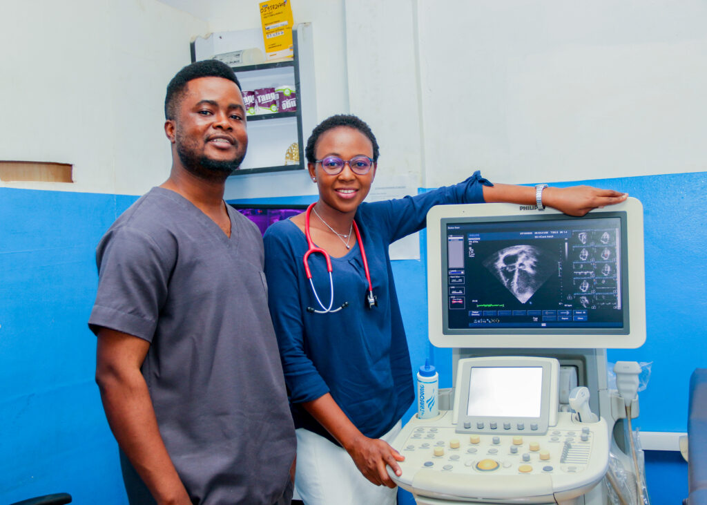
Ocular imaging, often referred to as eye scanning or ophthalmic imaging, involves various diagnostic techniques and technologies used to visualize and assess the structures of the eye. These imaging methods are essential for diagnosing and monitoring eye conditions, assessing vision problems, and guiding ophthalmic surgeries. Here are some common types of ocular imaging:
Fundus Photography: Fundus photography is a technique used to capture detailed images of the retina, optic nerve, and blood vessels in the back of the eye. These images are often used to monitor and diagnose conditions such as diabetic retinopathy, macular degeneration, and glaucoma.
Optical Coherence Tomography (OCT): OCT is a non-invasive imaging technique that provides cross-sectional images of the retina and other structures within the eye. It is invaluable for diagnosing and monitoring conditions like macular edema, retinal detachment, and glaucoma.
Fluorescein Angiography: Fluorescein angiography involves injecting a fluorescent dye into the bloodstream and taking rapid series of photographs to visualize the blood flow in the retina and choroid. It is useful for diagnosing retinal vascular conditions and identifying areas of leakage.
Indocyanine Green Angiography (ICG): ICG angiography is similar to fluorescein angiography but uses a different dye to visualize deeper structures within the eye, particularly the choroid. It is used in the evaluation of conditions like choroidal neovascularization.
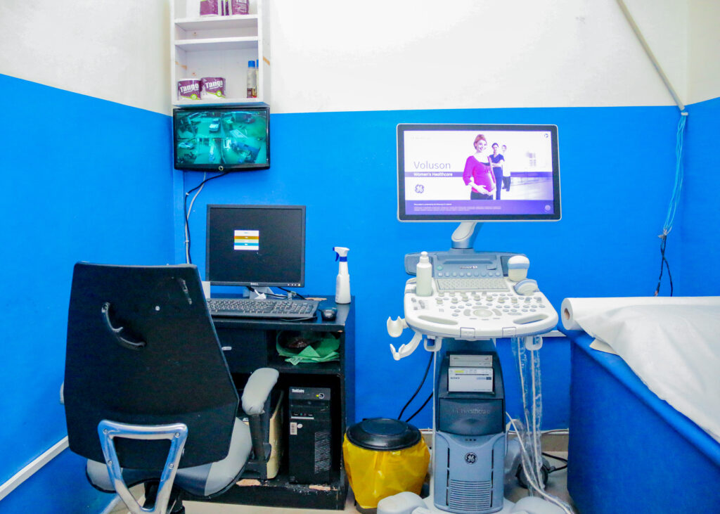
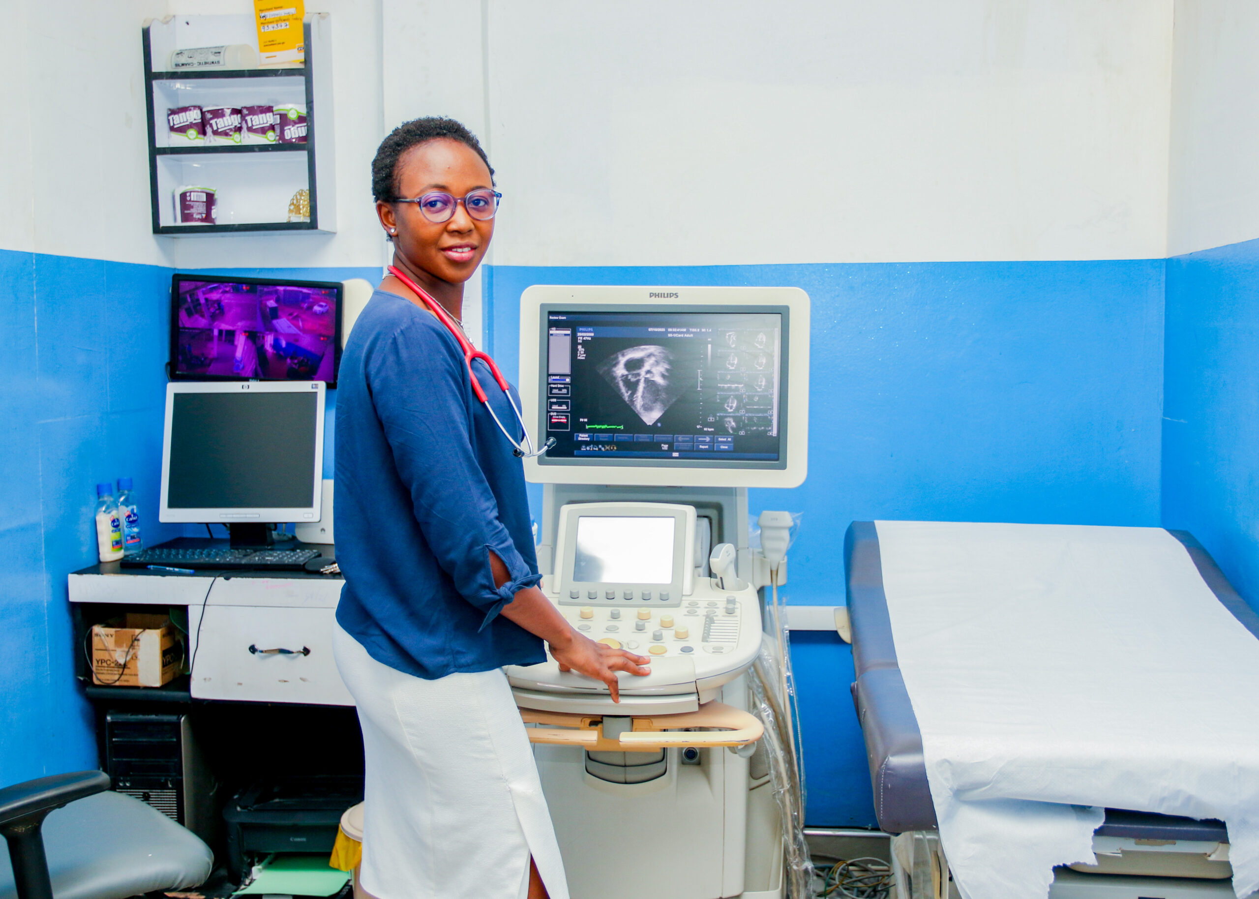
Ultrasound (Ocular Ultrasound): Ocular ultrasound is used to assess the eye’s internal structures, especially when visualization is challenging due to opaque media (e.g., cataracts or vitreous hemorrhage). It can help in diagnosing retinal detachments, tumors, and eye trauma.
Corneal Topography: Corneal topography measures the shape and curvature of the cornea, helping diagnose conditions like keratoconus and astigmatism. It’s essential for planning refractive surgeries like LASIK.
Pachymetry: Pachymetry measures the thickness of the cornea. This is important for diagnosing glaucoma and assessing corneal health.
B-scan Ultrasonography: B-scan ultrasound is used to evaluate the entire eye, including the posterior segment. It’s particularly useful for diagnosing conditions affecting the vitreous humor, retina, and posterior eye structures.
Anterior Segment Imaging: Techniques like anterior segment optical coherence tomography (AS-OCT) and slit-lamp photography are used to visualize the front part of the eye, including the cornea, iris, and lens. They help diagnose conditions like corneal dystrophies, cataracts, and glaucoma.
Visual Field Testing: While not imaging in the traditional sense, visual field testing assesses a person’s peripheral and central vision. It is valuable for diagnosing and monitoring conditions like glaucoma and neurological disorders affecting vision.
Ocular imaging is an essential component of ophthalmology and optometry. It allows eye care professionals to diagnose eye conditions, plan treatments, and track changes in eye health over time. Early detection and accurate diagnosis through ocular imaging can lead to better management of eye diseases and preservation of vision.
Our personal motivation consists of enhancing and improving patients’ experience in a sustainable manner. This is captured in our motto/slogan: ‘Legacy for Excellence’.


Copyright 2023 All Rights Reserved